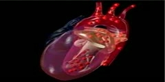
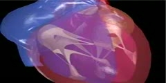
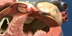
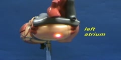
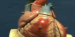
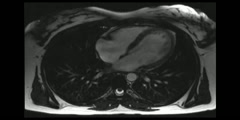
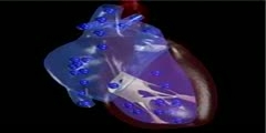
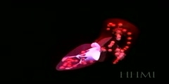
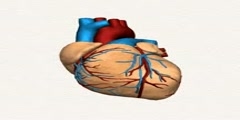
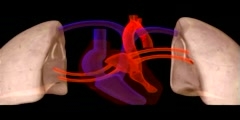
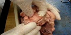
View of left heart valves
The mitral valve (so called because it resembles a bishop's miter) separates the left atrium from the left ventricle. The valve consists of two cusps of thin fibrous tissue, which form a hinge at the opening of the ventricle. As the ventricle fills with blood, the two cusps are pushed together by the increasing pressure inside the ventricle. At the other end of the valve, thin inelastic tendons (called chordae tendineae) attach the valve to the fingerlike papillary muscles of the ventricle. These attachments prevent the cusps of the valve from folding back into the atrium when the valve closes. Blood rushing into the empty ventricle from the atrium causes the valve cusps to open, and the cycle begins once again./nSemilunar valves get their name from their crescent moon shape. The aortic valve in the left heart is one of two semilunar valves; the other one is the pulmonary valve located in the right heart. It regulates blood flow from the left ventricle into the aorta. Its design is quite simple and effective: the valve is made up of three small cusps attached to a ring at the base of the aorta. When the ventricle is full of blood, the pressure causes each cusp to become "pinched" toward the wall of the aorta, and this in turn causes the valve to open. When the pressure in the ventricle drops, each cusp opens up again and the valve closes. This prevents blood in the aorta from flowing back into the ventricle.
