E. coli Infection mechanism
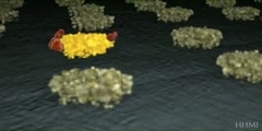
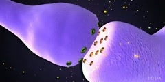
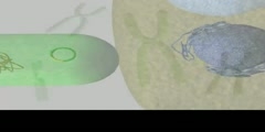
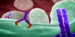
Watch this animation to see the molecular tricks that an infectious strain of Escherichia coli uses to infect your gut. Watch this three-part animation to see the molecular tricks that an infectious strain of Escherichia coli uses to infect your gut. E. coli are common, generally harmless bacteria, but certain less-common strains of E. coli can cause serious illness. An E. coli strain that's capable of infecting the intestinal tract is called enteropathogenic E. coli. Infection by enteropathogenic E. coli can cause severe diarrhea and can even result in death./nPart 1: A bacterium latches on to the surface of an intestinal cell/nThe surface of epithelial cells of the intestine is covered with microvilli, finger-shaped extensions of the cell that vastly increase the surface area for absorbing nutrients. In the animation, a single E. coli bacterium (purple) latches on to the surface of an intestinal epithelial cell (brown) using long, tetherlike pili. Pili are made of strands of long protein filaments that can adhere to the microvilli on the surface of intestinal cells./nOnce in contact with the bacterium, the microvilli disappear from a patch of the cell surface, the bacterium comes into closer contact with the intestinal cell surface, and the next phase in the infection process begins./nPart 2: The bacterium injects receptor proteins into the intestinal cell/nThe tethered bacterium now uses a specialized injector system to deliver some of its own proteins into the cell that it is invading. The injector systems that bacteria use are fascinating and are composed of several different proteins. In this case a Type III injector system is used, which is specialized for pumping things into other cells. The bacterium uses the injector system, much like a syringe, to introduce several bacterial proteins into the intestinal cell that force it to cooperate in its own infection./nA needlelike tube (purple) called EspA projects from the bacterium to the intestinal cell surface. Now two proteins (green) named EspB and EspD travel through the tube to form an opening in the intestinal membrane through which additional bacterial proteins can move into the cell. With the tube and pore complete, the bacterium now injects a protein called Tir (red) into the cell. The Tir proteins insert themselves into the intestinal cell membrane. A portion of the Tir protein projects beyond the cell surface and binds to a protein on the bacterial cell surface called intimin (blue cups). Now the membranes of the intestinal cell and the bacterium are locked together, and the intestinal cell is in big trouble. The Tir proteins become phosphorylated by intestinal cell proteins (blue balls). The stage is set for the next step, pedestal formation./nPart 3: Intestinal cell forms pedestal for bacterium, and infection follows/nThe bacterium is now firmly bound to the intestinal cell surface via the interaction between the Tir and intimin proteins. Pedestal formation, a very active and striking process, begins. Another intestinal cytoskeletal protein (orange booties) binds to a portion of the bacterial Tir protein that is inside the cell. Once these proteins bind, long strands of actin (yellow balls) start to form. The actin filaments build up directly beneath where the bacterium is bound to the intestinal cell. As the actin filaments lengthen, they push the cell membrane upward, and the bacterium becomes perched atop a dramatic pedestal formed by the intestinal cell./nOnce many enteropathogenic bacteria have adhered to the intestinal lining, symptoms of the infection (diarrhea) commence.
Channels: Cell Biology Microbiology
Tags: Ecoli Infection mechanism
Uploaded by: admin ( Send Message ) on 12-04-2012.
Duration: 2m 52s
