Structure and Function of Cytochrome
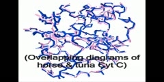
This animation shows an 'inside view' of the workings of the enzyme cytochrome P-450 2C9. The protein, represented by the ribbon and yellow spheres, is from an x-ray crystal structure of a drug metabolizing enzyme called cytochrome P-450 2C9. The larger of the two ligands (clusters of spheres) is the heme group, which acts as cofactor to assist in the catalytic reaction. The smaller of the two ligands is the drug warfarin (an anticoagulant) which is the substrate for the catalytic reaction. First both ligands are bound. Then the warfarin molecule moves from solution (outside the protein) and finds a channel by which to access its specific binding site. The warfarin molecule finds its way through the channel to find its preferred binding position near the active site.This model will now be used to illustrate how different drugs interact with this enzyme, and thus interfere with optimum warfarin therapy, a common clinical problem.Created by:Steven Barbera - Business/CommunicationsSteven BarberaClass of 2007Business/CommunicationsSTA member since 2003Dr. King - Biomedical and Pharmaceutical SciencesDr. Roberta KingAssistant Professor of Biomedical and Pharmaceutical SciencesCollege of Pharmacy
Channels: Scientific Animations Biochemistry
Tags: cytochrome
Uploaded by: watchme ( Send Message ) on 04-11-2007.
Duration: 0m 45s
