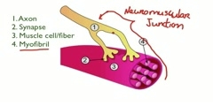
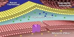
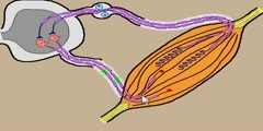
Skeletal Muscle Fiber Model - Neuromuscular Junction
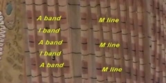
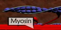
Skeletal Muscle Fiber Structure A myofiber is a multinucleated single muscle cell (see Figure 1, below). Physically, they range in size from a under a hundred microns in diameter and a few millimeters in length to a few hundred microns across and a few centimeters in length. The cell is densely packed with contractile proteins, energy stores and signaling mechanisms. Figure 1: Myonuclei identified along the length of an isolated muscle fiber. Nuclei were stained with a florescent dye that binds to DNA. (Micrograph kindly provided by V. Reggie Edgerton (University of California, Los Angeles).
Channels: Scientific Animations
Tags: Skeletal Muscle Fiber Model Neuromuscular Junction Metabolism Excitation
Uploaded by: buraktube ( Send Message ) on 09-11-2010.
Duration: 3m 16s
