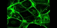
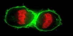
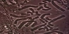
Cell divison video under flourescent microscopy
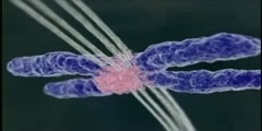
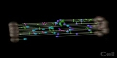
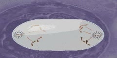
This video shows the beauty of spindle during cell division. As it is known, during cell division, microtubules are formed and these microtubules are attached to kinetechore region of chromosomes. Kinetechore is a special DNA sequence region. When microtubules binds to kinetechore, it seperated chromosomes from each other. As you see in the video, chromosomes are about to be seperated. Blue one is DAPI staining showing the presence of DNA. Red color shows the location of kinetechore. And green color shows microtubules.
Channels: Scientific Animations Cell Biology Genetics Medical
Tags: mitotic spindle human cells division microtubules chromosomes mithosis fluorescent dye
Uploaded by: tubeman ( Send Message ) on 30-03-2007.
Duration: 0m 31s
