Lifecycle of an miRNA ( microRNA)
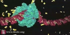
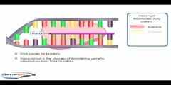
This 3D animation provided courtesy of Rosetta Genomics shows how microRNA works. MicroRNAs (miRNA) are single-stranded RNA molecules of about 21-23 nucleotides in length thought to regulate the expression of other genes. miRNAs are encoded by genes that are transcribed from DNA but not translated into protein (non-coding RNA); instead they are processed from primary transcripts known as pri-miRNA to short stem-loop structures called pre-miRNA and finally to functional miRNA. Mature miRNA molecules are partially complementary to one or more messenger RNA (mRNA) molecules, and they function to downregulate gene expression. More info: The genes encoding miRNAs are much longer than the processed miRNA molecule; miRNAs are first transcribed as primary transcripts or pri-miRNA and processed to short, 70-nucleotide stem-loop structures known as pre-miRNA in the cell nucleus. This processing is performed in animals by a protein complex known as the Microprocessor complex, consisting of the nuclease Drosha and the double-stranded RNA binding protein Pasha.[2] These pre-miRNAs are then processed to mature miRNAs in the cytoplasm by interaction with the endonuclease Dicer, which also initiates the formation of the RNA-induced silencing complex (RISC).[3] This complex is responsible for the gene silencing observed due to miRNA expression and RNA interference. The pathway in plants varies slightly due to their lack of Drosha homologs; instead, Dicer homologs alone effect several processing steps.[4] Zeng et al. have shown that efficient processing of pre-miRNA by Drosha requires presence of extended single-stranded RNA on both 3'- and 5'-ends of hairpin molecule. They demonstrated that these motifs could be of different composition while their length is of high importance if processing is to take place at all. Their findings were confirmed in another work by Han et al. Using bioinformatical tools Han et al. analysed folding of 321 human and 68 fly pri-miRNAs. 280 human and 55 fly pri-miRNAs were selected for further study, excluding those molecules which folding showed presence of multiple loops. All human and fly pri-miRNA contained very similar structural regions, which authors called 'basal segments', 'lower stem', 'upper stem' and 'terminal loop'. Based on the encoding position of miRNA, i.e. in the 5'-strand (5'-donors) or 3'-strand (3'-donors), thermodynamical profiles of pri-miRNA was determined. Following experiments have shown that Drosha complex cleaves RNA molecule ~2 helical turns away from the terminal loop and ~1 turn away from basal segments. In most analysed molecules this region contain unpaired nucleotides and the free energy of the duplex is relatively high compared to lower and upper stem regions. Most pre-miRNAs don't have a perfect double-stranded RNA (dsRNA) structure topped by a terminal loop. There are few possible explanations for such selectivity. One could be that dsRNAs longer than 11 base pairs activate interferon response and anti-viral machinery in the cell. Another plausible explanation could be that thermodynamical profile of pre-miRNA determines which strand will be incorporated into Dicer complex. Indeed, aforementioned study by Han et al. demonstrated very clear similarities between pri-miRNAs encoded in respective (5'- or 3'-) strands. When Dicer cleaves the pre-miRNA stem-loop, two complementary short RNA molecules are formed, but only one is integrated into the RISC complex. This strand is known as the guide strand and is selected by the argonaute protein, the catalytically active RNase in the RISC complex, on the basis of the stability of the 5' end.[5] The remaining strand, known as the anti-guide or passenger strand, is degraded as a RISC complex substrate.[6] After integration into the active RISC complex, miRNAs base pair with their complementary mRNA molecules and induce mRNA degradation by argonaute proteins, the catalytically active members of the RISC complex. It is as yet unclear how the activated RISC complex locates the mRNA targets in the cell, though it has been shown that the process is not coupled to ongoing protein translation from the mRNA.[7] REf: http://en.wikipedia.org/wiki/Micro_RNA
Channels: Scientific Animations Genetics Molecular Biology
Tags: DNA RNA transcription micro RNA
Uploaded by: benchwork ( Send Message ) on 26-03-2007.
Duration: 0m 46s

